I. INTRODUCTION
Shanghai Synchrotron Radiation Facility (SSRF) is a third-generation synchrotron radiation source with a 3.5 GeV storage ring [1]. At present, it is operated in a top-up injection mode with a constant electron beam current of 240 mA. The beamline BL16B1 at SSRF was designed for measurements of small angle X-ray scattering (SAXS), wide angle X-ray scattering (WAXS), anomalous small angle X-ray scattering (ASAXS), and grazing incident small angle X-ray scattering (GISAXS). Constructed and opened to users in 2009, and upgraded in 2013, the beamline enables time-resolved studies on structural transitions in the sub-second level. Most users’ projects performed on this beamline refer to kinetics, dynamics and rheology studies on materials with nanoscale structures, such as polymers, nanomaterials, mesoporous materials, colloids, liquid crystals, metal materials, etc. In this paper, we present a detail description about the SAXS beamline and station.
II. BEAMLINE CONFIGURATION AND TECHNIQUE SPECIFICATIONS
The photon source of bending magnet delivers 5 keV to 20 keV X-rays. The beam with 1.2×0.12 mrad2 divergence can be accepted by the optics system. The beamline optics consists of a Si(111) flat double crystal monochromator (DCM) and a double focusing toroidal mirror. Optical layout of the beamline is shown in Fig. 1. Generally, the beamline works at 10 keV (0.124 nm) for user experiments.

Specifications of the SAXS beamline are as follows:
Energy range: 5–20 keV;
Energy resolution: 4.0×10⁻⁴@10 keV;
Flux: ∼3×10¹¹ phs/s@10 keV and 240 mA;
Focus size: 0.4 mm (H)× 0.5 mm (V).
The results of spot size and rocking curve measured on BL16B1 are shown in Fig. 2.

The endstation of BL16B1 is shown in Fig. 3. The detectors are Mar165 CCD for SAXS and INEL CPS120, a one-dimensional arc gas detector, for WAXS. The beam intensity monitor before sample is a N2 gas ionization chamber, and the monitor after sample adsorption is a photodiode in the beam stop. Two scatter-less slits (Xenocs) are used to depress parasitic scattering. The sample holder is mounted onto an optical table. The distance of sample to SAXS detector can be adjusted up to 5 m, and scattering from a sample transmits in vacuum and passes Kapton window to hit the detector. There are sample stages for SAXS, GISAXS or WAXS (2D). The q ranges are 0.03–3.6 nm-1 for SAXS measurement and 4.5–33 nm-1 for WAXS measurement at 10 keV. And a continuous q range of 0.06–33 nm-1 can be obtained in simultaneous SAXS/WAXS. Two conventional modes in sample-to-director distance of 2 m and 5 m have been provided to users, with the q ranges being 0.08–3.6 nm-1 at 2 m, and 0.03–1.4 nm-1 at 5 m.
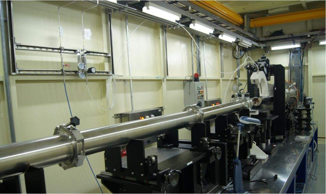
The beamline control system works on an EPICS platform in Linux operation system, which is convenient to communicate among the devices; though the CPS 120 detector works currently on a Labview software platform in Windows operation system (it will be integrated into EPICS in future). Besides the foreseen sample surroundings, the users have the possibility to install their own specialized sample devices [2-5]. Besides technical boundary conditions, user friendliness and reliability have been considered as important criteria.
III. METHODOLOGY
Techniques of the beamline include SAXS, WAXS, simultaneous SAXS/WAXS, GISAXS and ASAXS.
A. SAXS technique
SAXS in transmission mode can be used for materials studies, such as polymer, nanomaterials, metal, bio-materials, etc. [6-10]. Most of the SAXS experiments finished are in-situ studies on microstructure evolution of different samples, such as in-situ stretching of polymers and fiber at different temperatures. A typical work [11] is described as follows.
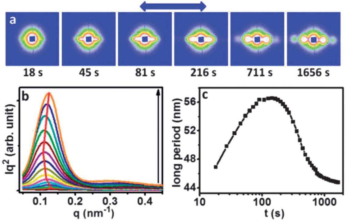
The long time evolution of extension-induced crystallization of polyethylene oxide (PEO) was investigated with a combination of rheological measurement and in-situ SAXS, aimed at understanding dynamic changes in spatial arrangement of nuclei, in terms of both the chain stretching and orientation. The structural evolution in the small strain region is shown in Fig. 4.
B. Simultaneous SAXS/WAXS measurement
Figure 5(a) shows the simultaneous SAXS/WAXS system. In order to obtain a continuous scattering vector q across the SAXS and WAXS collectable regions, the simultaneous SAXS/WAXS technique was developed, with the determinable range of SAXS measurement being q= 0.06–3.50 nm-1, while that of WAXS being q= 2.50–34.8 nm-1. Therefore, a continuous q can be obtained with a continuous q range of 0.06–33 nm-1 (Fig. 5(b)). Simultaneous SAXS/WAXS is an important tool to study fiber stretching, liquid crystal and self-assembly system [12, 13].
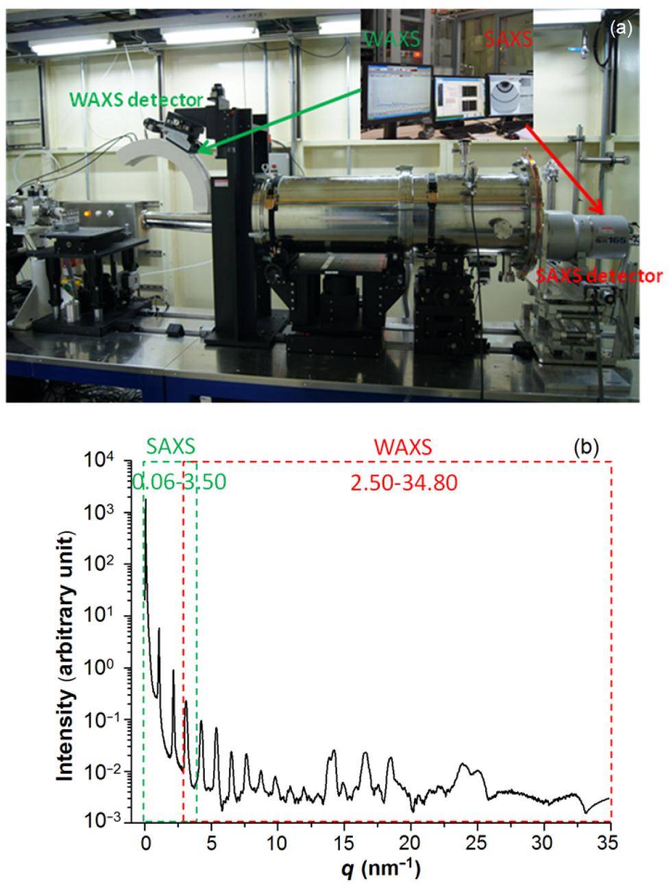
The hardware is shown schematically in Fig. 6. The Mar165 CCD is the SAXS detector and the 1D arc PSD (CPS 120) is the WAXS detector. The SAXS detector has a trigger signal to control other equipment to work synchronously. The shutter, and WAXS and SAXS detectors, can work synchronously through a pulse generator. Simultaneous SAXS/WAXS results for silver Behenate performed on BL16B1 are shown in Fig. 5(b). The SAXS and WAXS data are combined through scaling the SAXS data with a factor to equal their integral area in the overlap region.

C. Grazing incidence SAXS
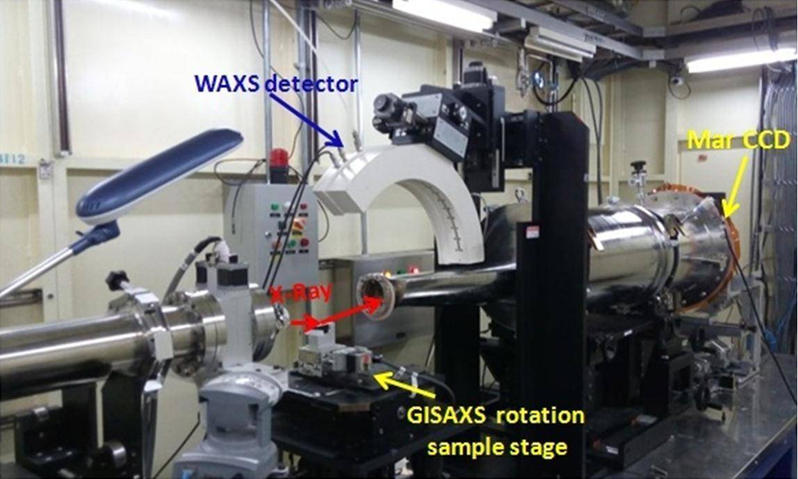
GISAXS method is a powerful tool for studying film surface and interface structures [14-17]. A Kohzu tilt stage is used as GISAXS sample stage (Fig. 7). The incidence angle of X-rays can be adjusted by the sample stage with an accuracy of 0.001. In principle, the size of samples should be larger than 0.5 cm (w)× 1 cm (l).
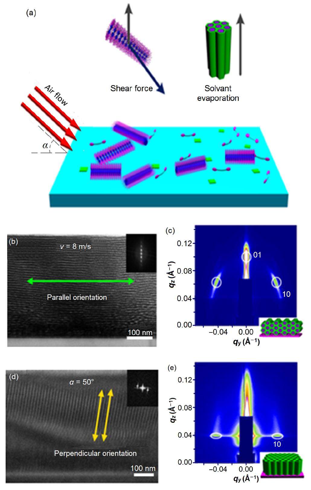
The GISAXS studies [18-21] performed on BL16B1 include the finding of a novel orientation of mesochannels in mesostructured thin films, and the results were published in Journal of American Chemical Society [22].
As shown in Fig. 8(a), uniaxially oriented mesoporous titania films were prepared in any uniaxial alignment direction by manipulating the magnitude and incidence angle of a shear-force of a hot air-flow. According to TEM and GISAXS results (Figs. 8(b)–8(e), orientation of the mesochannels can be well controlled into parallel, vertical or oblique, with any angle with respect to the plane, by simply regulating the rate and incident angle of the air flow.
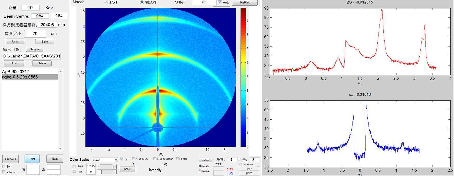
A one dimension profile along the vertical and horizontal directions can be obtained quickly from a two dimension GISAXS pattern using "Pre-GISAXS" (Fig. 9) [23].
D. Anomalous SAXS
Depending on energy tunability, ASAXS can be conducted. ASAXS of aluminum alloys (7150 and 7085 Al alloy) has been carried out. The SAXS patterns of 7150 Al alloy were obtained at various energies below the Zn K-edge. It was found that the scattering intensity increased obviously with decreasing energy. Due to absorption, thickness of the alloy sample should be less than 150 μm.
E. SAXS study of protein solution
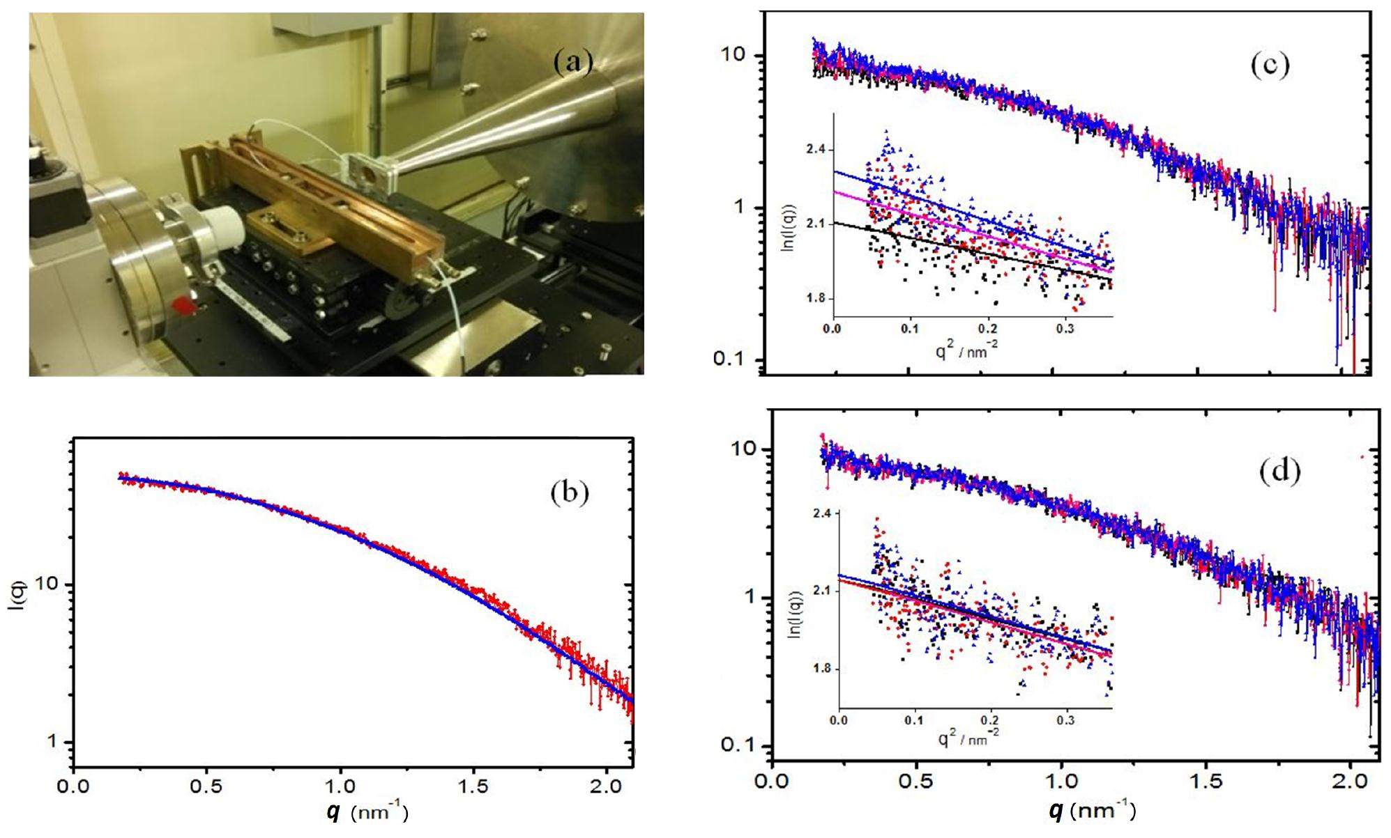
To reduce radiation damage, we have developed a peristaltic device with thermo controller for solution SAXS measurements (Fig. 10(a)) [24]. Figures 10(b)–10(c) show the scattering data of lysozyme collected at BL16B1 using the device, indicating that the device is able to reduce the radiation damage for biomacromolecule solution samples.
F. Supporting conditions and data treatment
The BL16B1 beamline at SSRF offers support laboratories for experimental research of users. Sample preparation can be performed in support laboratories with ultra-pure water system, magnetic stirrer, electronic balance, ultrasonic apparatus, centrifuge, electric oven, vacuum drying apparatus, polarizing microscope and AFM. And some in-situ devices are available in this beamline, such as Linkam THMS600 heating stage (-196 ℃ to 600 ℃), Linkam CSS450 shearing system (ambient to 450 ℃), Linkam TS1500 heating stage (ambient to 1500 ℃), in-situ fiber stretching (maximum tension: 1000 N) device (ambient to 500 ℃), and helium-atmosphere sample chamber (to protect the samples from oxidation).
A few widely-used packages of data treatment have been developed, such as Irena, Nika, X-polar, FIT2D, etc.
IV. CONCLUSION
The SAXS beamline at SSRF provides powerful support in studying microstructure of polymers, fibers and nanostructure/mesoporous materials. The techniques available in the beamline include SAXS, WAXS, ASAXS, GISAXS, and time-resolved simultaneous SAXS/WAXS studies on structural transitions in the sub-second time region and partly ordered systems with a scaling of 1 to 240 nm in real-space. More techniques will be developed at BL16B1, such as USAXS technique based on Bonse-Hart camera system and micro-focus SAXS. Still more common in-situ devices to facilitate the users will be developed. A complete SAXS data analysis platform will be established. This platform will include data pretreatment, and SAXS and WAXS data analysis.
Current status and progresses of SSRF project
. Nucl Sci Tech, 2008, 19: 1-6. DOI: 10.1016/S1001-8042(08)60013-5Critical stress for crystal transition in poly(butylene succinate)-based crystalline-amorphous multiblock copolymers
. Macromolecules, 2014, 47: 7533-7539. DOI: 10.1021/ma501832zStudy of the influence of TiB2 particles on the melt structure of the hypoeutectic Al-Cu alloy by small angle X-ray scattering
. Mater Chem Phys, 2014, 143: 471-475. DOI: 10.1016/j.matchemphys.2013.10.027Synchronous stimuli of biodegradable poly(butylene succinate-co-terephthalate) copolymer via uniaxial stretching at varying temperatures
. J Polym Sci Pol Phys, 2014, 9: 640-649. DOI: 10.1002/polb.23681Self-reinforced polyethylene blend for artificial joint application
. J Mater Chem B, 2014, 2: 971-980. DOI: 10.1039/C3TB21231ANanoconfined crystallites toughen artificial silk
. J Mater Chem B, 2014, 2: 1408-1414. DOI: 10.1039/C3TB21148GDynamic and quantitative control of the DNA-mediated growth of gold plasmonic nanostructures
. Angew Chem Int Edit, 2014, 32: 8338-8342. DOI: 10.1002/anie.201402937In situ studies of microstructure evolution and properties of an Al-7.5Zn-1.7Mg-1.4Cu-0.12Zr alloy during retrogression and reaging
. Mater Design, 2014, 56: 1020-1024. DOI: 10.1016/j.matdes.2013.12.006Amphiphilic starlike dextran wrapped superparamagnetic iron oxide nanoparticleclsuters as effective magnetic resonance imaging probes
. Biomaterials, 2013, 34: 1193-1203. DOI: 10.1016/j.biomaterials.2012.10.056Skeletal cubic, lamellar, and ribbon phases of bundled thermotropic bolapolyphiles
. J Am Chem Soc, 2014, 136: 6846-6849. DOI: 10.1021/ja502410eRelaxation propelled long period change in the extension induced crystallization of polyethylene oxide
. Soft Matter, 2013, 9: 10759-10767. DOI: 10.1039/C3SM52152DPhase diagrams of stoichiometric polyelectrolyte-surfactant complexes
. Macromolecules, 2003, 36: 9549-9558. DOI: 10.1021/ma034352cSelf-assembled hierarchical structure evolution of azobenzene-containing linear-dendritic liquid crystalline block copolymers
. Soft Matter, 2012, 8: 6174-6184. DOI: 10.1039/C2SM07249ALamellar diblock copolymer thin films during solvent vapor annealing studied by GISAXS: different behavior of parallel and perpendicular lamellae
. Macromolecules, 2014, 47: 5711-5718. DOI: 10.1021/ma500633bSmall- and wide-angle X-ray scattering characterization of bulk heterojunction polymer solar cells with different fullerene derivatives
. J Phys Chem C, 2012, 116: 10238-10244. DOI: 10.1021/jp210140jHierarchical structure in nanoscale thin films of a poly(styrene-b-methacrylate grafted with POSS) (PS214-b-PMAPOSS27)
. Macromolecules, 2010, 43: 10568-10581. DOI: 10.1021/ma101276dRegioregular oligomer and polymer containing thieno[3,4-b]thiophene moiety for efficient organic solar cells
. Macromolecules, 2009, 42: 1091-1098. DOI: 10.1021/ma8023969Systematic investigation of side-chain branching position effect on electron carrier mobility in conjugated polymers
. Adv Funct Mater, 2014, 40: 6270-6278. DOI: 10.1002/adfm.201401822Airflow-field-induced sandwich-type membrane of block copolymer for selective ion separation
. Macromol Rapid Comm, 2014, 35: 735-740. DOI: 10.1002/marc.201300880Contamination-resistant silica antireflective coating with closed ordered mesopores
. Phys Chem Chem Phys, 2014, 16: 16684-16693. DOI: 10.1039/c4cp01032aSynthesis and characterization of room temperature columnar mesogens of cyclotriphosphazene with Schiff base units
. J Mater Chem C, 2013, 1: 7148-7154. DOI: 10.1039/C3TC31371AA facile approach for controlling the orientation of one-dimensional mesochannels in mesoporous titania films
. J Am Chem Soc, 2012, 134: 20238-20241. DOI: 10.1021/ja309168fCalibrating scattering angle in wide angle X-ray scattering experiment
. Sci China Ser G-Phys Mech Astron, 2015, 45: 017001. (in Chinese) DOI: 10.1360/SSPMA2014-00106The resource for macromolecular SAXS
. http://www.bioisis.net/tutorial
